LINEO TUTORIAL

NOTES OF ORTHODONTICS

THE BIOMECHANICAL MOVEMENTS

The movement of teeth during orthodontic treatment causes reactions and changes that affect the periodontium, alveolar bone, dentin, and cementum. Orthodontic movement is the result of the biological response of these structures to the forces applied by mechanical systems. This response is characterized by a cascade of events triggered by compression/tension of the periodontal ligament, which leads to an inflammatory process underlying tissue remodeling, generating resorption and neoapposition of bone tissue. The intensity of the forces and how they are transmitted to the teeth, and thus to the periodontium, are crucial in determining the type of tissue response. It is known that continuous forces give better biological responses with faster and safer movements to the periodontium and tooth roots. In addition, forces must also have a threshold value; below this value, the periodontal ligament has the ability to stabilize the tooth through active metabolism.

In orthodontics, biomechanics represents the application of mechanics to the biology of tooth displacement and is based on certain principles
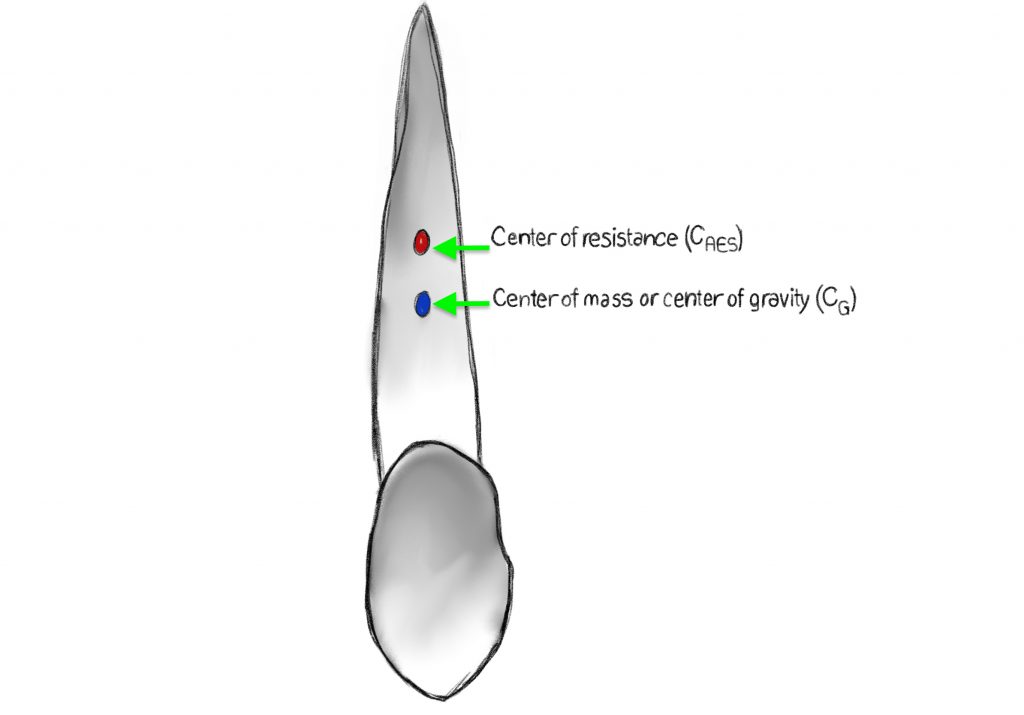
Center of Mass: A tooth, like any other body, has a point in its mass located at its center. If you apply a force directly on this point, the tooth will move in a linear direction, without rotation. In practice, it will move in a bodily manner, translating, that is, the crown and the root will move in the same direction and by the same amount of space.

Center of Resistance (CRES) : the tooth is located within the alveolar bone, its displacement is restricted and its center moves apically. It also depends on the amount of alveolar bone and the length of the root.
Center of Rotation: represents the point around which the same body rotates following the application of a force; its position will determine the type of dental movement.
Torque: represents the measure of the rotational tendency produced by that force. If the force is applied at any point other than the center of resistance, the tooth will move linearly but with a rotation, so its axial inclination will change. In orthodontics, it is impossible to apply a single force that passes through the center of resistance so a torque is inevitably created.

TOOTH MOVEMENTS

Six types of tooth movement can be identified based on where the center of rotation is in relation to the CRES.
Uncontrolled tipping: occurs when, by applying a force on the crown of a tooth, it moves in the direction of the applied force while the root moves in the opposite direction. The center of rotation is anywhere between the center of resistance and the root apex.
Controlled tipping: movement similar to uncontrolled tipping but the center of rotation is near or coincident with the root apex (the tooth moves like a clock pendulum, with the apex corresponding to the center of rotation and the crown moving from side to side) Note that in biomechanics this is referred to as tipping.
Root movement: the center of rotation is located at the top of the crown while the root is free to move in the direction of the applied force. It is also defined as root “torque” i.e. with root displacement and with no or minimal crown displacement. From a biomechanical point of view, the movement is similar to controlled tipping.
Pure translation: both the crown and the root move in the same direction and by the same amount of space without rotation. In this case the center of rotation is nonexistent or, in mathematical terms, approaches infinity.
Pure extrusion and intrusion: two types of vertical movements in which the former represents the movement the tooth makes when moving in the same direction as its eruption, while intrusion is the movement of the tooth toward the alveolar bone.
Pure rotation: means the movement of a tooth around the center of rotation and the end of the crown and root move in opposite directions, describing an arc of a circle.

FORCE SYSTEMS

Clinically, forces can be applied in three ways to determine tooth displacement:

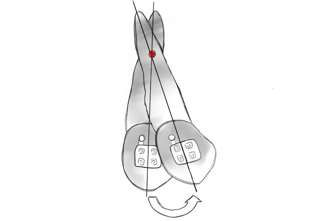
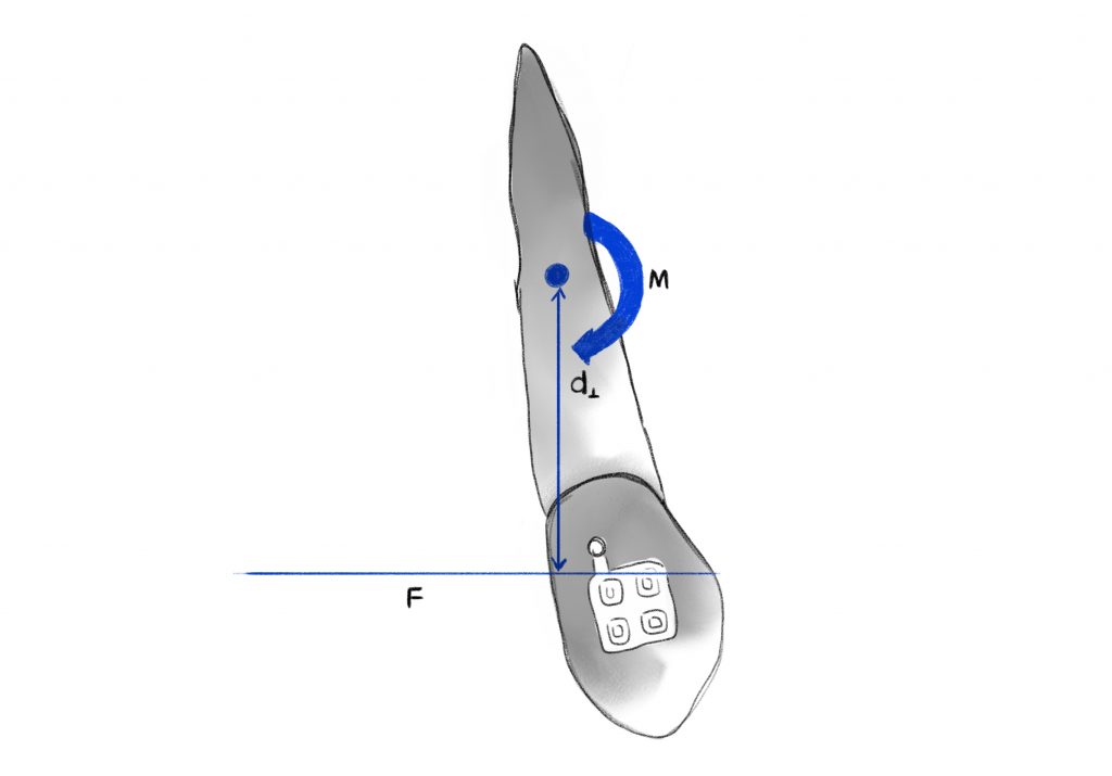
1.Single force that results in significant crown inclination;

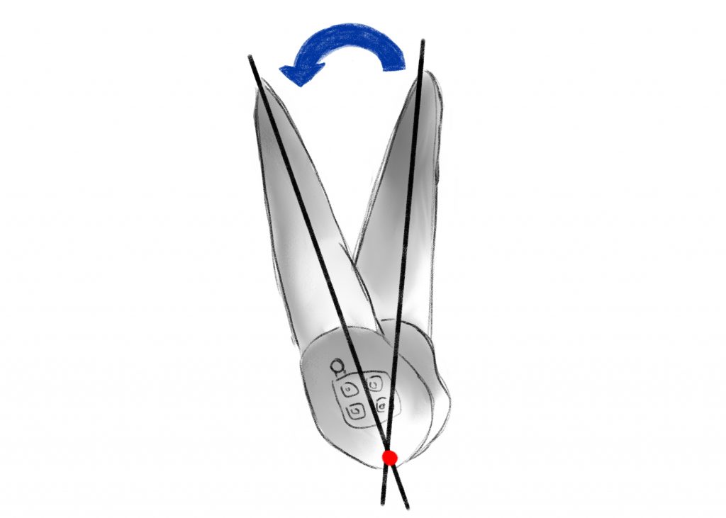
2.Pair of forces that allows a controlled inclination of the root in the mesio-distal sense (tipping) or in the vestibulo-lingual sense (torque);

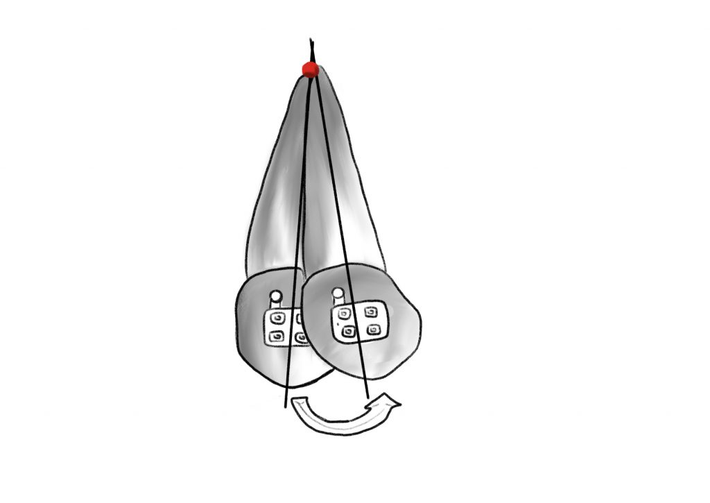
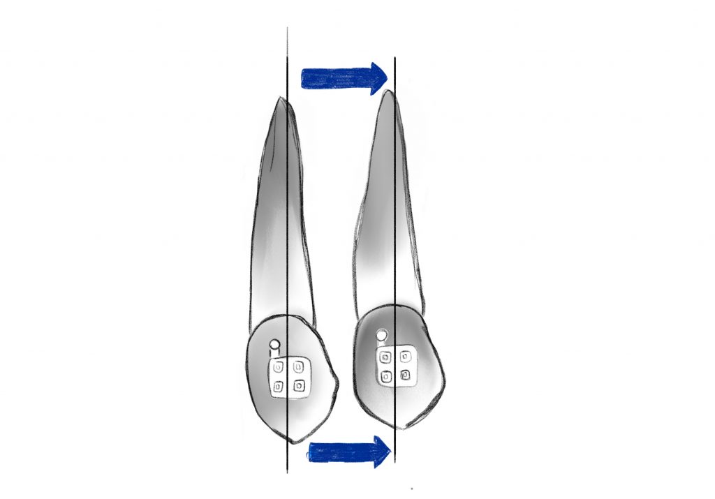
3.Single force + force pair, a combination that allows to obtain a translation of the tooth while maintaining good control of the root inclination.

Bibliografia

1.      Burstone CJ, Pryputniewicz RJ. Holographic determination of centers of rotation produced by orthodontic forces. Am J Orthod. 1980;77:396.

2.      Davidian EJ. Use of a computer model to study the force distribution on the root of the maxillary central incisor. Am J Orthod. 1971;59:581–588.

3.      Hay GE. The equilibrium of a thin compressible membrane. Can J Res. 1939;17:106–121.

4.      Yettram AL, Wright KWJ, Houston WJB. Center of rotation of a maxillary central incisor under orthodontic loading. Br J Orthod. 1977;4:23–27.

5.      Christiansen RL, Burstone CJ. Centers of rotation within the periodontal space. Am J Orthod. 1969;55:351–369.

6.      Smith RJ, Burstone CJ. Mechanics of tooth movement. Am J Orthod.1984;85(4):294–307.

7.      Pryputniewicz RJ, Burstone CJ. The effect of time and force magnitude on orthodontic tooth movement. J Dent Res. 1979;58(8):1754–1764.

8.      Mulligan TF. Common sense mechanics in everyday orthodontics. Phoenix, AZ: CSM Publishing; 1998:1–17.

9.      Isaacson RJ, Lindauer SJ, Rubenstein LK. Activating a 2 × 4 appliance. Angle Orthod. 1993;63:17–24.

10.   Demange C. Equilibrium situations in bend force systems. Am J Orthod Dentofacial Orthop. 1990;98:333–339.

11.   Koenig HA, Burstone CJ. Force systems from an ideal arch. Am J Orthod. 1974;65:270–289.

12.   Burstone CJ, Koenig HA. Force systems from an ideal arch: large deflection considerations. Angle Orthod. 1989;59(1):11–16.

13.   Koenig HA, Burstone CJ. Creative wire bending: the force system from step and V bend. Am J Orthod Dentofacial Orthop. 1988;93(1):59–67.

MALOCCLUSIONS

Dental malocclusions are represented by an abnormal relationship between the teeth of the upper and lower jaws and can occur in the three planes of space (vertical, transverse, and sagittal). The etiology of dento-skeletal alterations is multifactorial, and three causative factors (hereditary, congenital, and acquired) can be identified. With regard to hereditary factors, the etiopathogenesis of malocclusions is polygenic and of variable expressivity so that, in general, the familial recurrence of an alteration is assessed.

Congenital factors are acquired intrauterine and can have repercussions in the oral cavity (medication, trauma and infectious diseases). Lastly, acquired factors are those that occur after birth, such as spoiled habits (thumb sucking, onychophagia, atypical swallowing, oral respiration), facial and/or dental traumas, inflammatory and neoplastic pathologies.

The presence of malocclusions can cause problems of various kinds:

Functional (disorders of phonation, chewing, swallowing or breathing);

Aesthetic, as they can alter the aesthetics of the smile;

Social and emotional, as severe malocclusions can cause psychological problems;

Articolari (disturbi dell’articolazione temporo-mandibolare);

Dental, as misaligned teeth can lead to an increased risk of tooth decay and periodontal disease.

The concept of “ideal”: The purpose of orthodontic treatment is to transform a malocclusion into an “ideal” dento-skeletal Class I occlusion. The word dento-skeletal indicates that an “ideal” occlusion consists of two components (dental and skeletal).

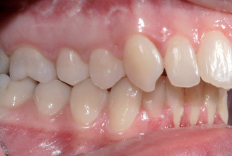
When the teeth are in occlusion, the mesio-vestibular cusp of the upper first molar occludes in the vestibular sulcus of the lower first molar (Angle’s Class I molar ratio), with the upper canines located between the canines and the lower first premolars (Class I canine ratio).

Other dental characteristics of an “ideal” occlusion are:

Maximum intercuspation;

Overjet (distance, measured parallel to the occlusal plane, between the vestibular surface of the lower incisor and the incisal margin of the upper incisor) with normal values from 0-2 mm;

Overbite (distance, measured perpendicular to the occlusal plane, between the incisal edge of the lower incisor and the incisal edge of the upper incisor) of 2-3 mm
The median lines of each arch coinciding with each other.

With regard to the skeletal component, it is essential to refer to the so-called skeletal “norm”, established by the dento-cranio-facial relationships of selected individuals with “harmonious” occlusions, in order to identify and quantify the deviations or disharmonies that each patient presents with respect to normal values and to establish the most appropriate treatment to determine those changes necessary to correct the disharmonies.

SAGITTAL PLANE MALOCCLUSIONS

Classification of malocclusions:

The classification according to Angle is based on the relationships that the upper teeth contract with the lower teeth in two areas (molar and canine). Malocclusions can be divided into three groups (Class I, II and III).

Class I malocclusions are defined as those in which the molars are in class I relationships and the incisors are crowded and/or protruded, in one or both arches.

A Class II malocclusion is defined by the position of the mesio-vestibular cusp of the upper first molar occluding mesial to the mesio-vestibular sulcus of the lower first molar or occluding head to head (Class II molar), and/or the lower canine occluding more distal than Class I (Class II canine). 
In addition, Class IIs are further divided into:

Class II division 1 (Class II occlusion with highly vestibularized incisors and increased overjet)

Class II division 2 (Class II occlusion with retroclined central incisors and often proclined lateral incisors)

Class II sub division (Class II occlusion on one side and Class I occlusion on the opposite side).

In Class III malocclusion, the mesio-vestibular cusp of the upper first molar occludes distal to the vestibulo-mesial sulcus of the lower first molar (Class III molar), and/or the lower canine occludes more mesial than Class I (Class III canine).

Class IIIs are further divided into: Class III true (Class III molar relationship in centric relation, is skeletal in nature with an overdeveloped mandible and/or hypereveloped maxilla) and pseudo-Class III (Class I malocclusion complicated by an anterior cross bite that causes forward closure of the mandible relative to its position in centric relation)

Bibliografia

1.      Petroviç D, Vukiç-Culafi B, Iviç S, Djuriç M, Milekiç B. Study of the risk factors associated with the development of malocclusion. Vojnosanit Pregl 2013;70(9):817-23.

2.      Bolton W.A. The clinical application of a tooth size analysis. Am J Orthod. 1962;48:504-529.

3.      Moorrees CFA, Chada JM. Available space for incisors during dental development: a growth study based on physiologic age. Angle Orthod 1965;35:12-22.

4.      Moorres CFA, Gron AM, Lebret LM, Yen PHJ, Frolich FJ. Growth studies of dentition. A review. Am J Orthod 1969;55:600-16.

5.      Enlow DH. Handbook of Facial Growth. (2nd ed) WB Saunders, Philadelphia 1985.


MALOCCLUSIONS ON THE TRANSVERSE PLANE

A fundamental prerequisite for achieving normal occlusal relationships during orthodontic treatment is the correct positioning in the three planes of space of the maxillary bone and mandible, which must also be correctly proportioned to each other.

Under normal conditions, the right and left halves of the face are almost symmetrical and the upper arch overlaps the lower arch for its entire length. When this condition does not occur, there is a “transverse discrepancy,” which represents a “developmental abnormality in the transverse plane.” Although the diagnosis of transverse discrepancy is often straightforward, some asymmetries can sometimes be masked by dental compensations. Depending on the structures involved, these problems may have a dental, skeletal, or functional origin, or a combination of these factors may be present.

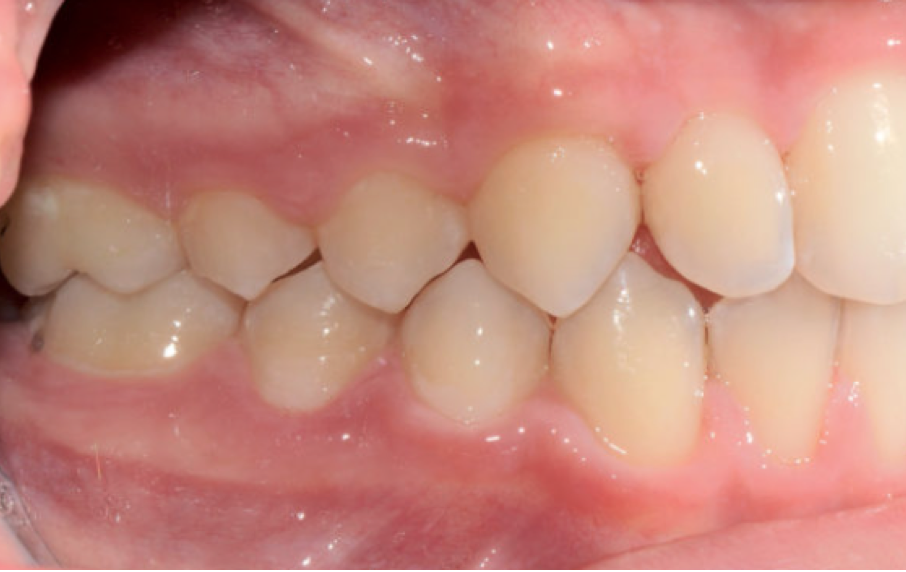
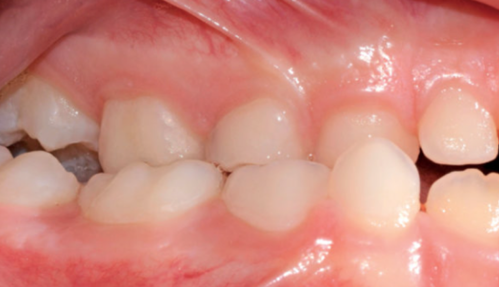
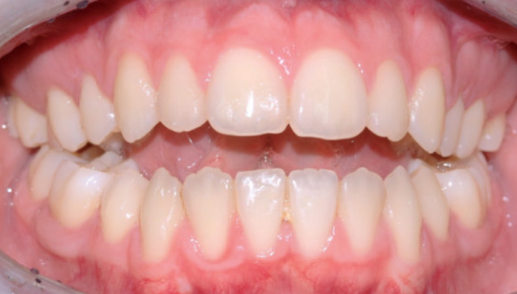
In a normal occlusion, in the transverse plane, the palatine cusps of the upper molars and premolars occlude into the pits of the lower molars and premolars while in the anteroposterior plane, the upper incisors occlude on the buccal surfaces of the lower incisors.

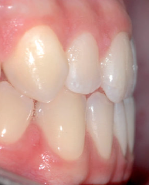
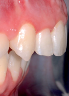
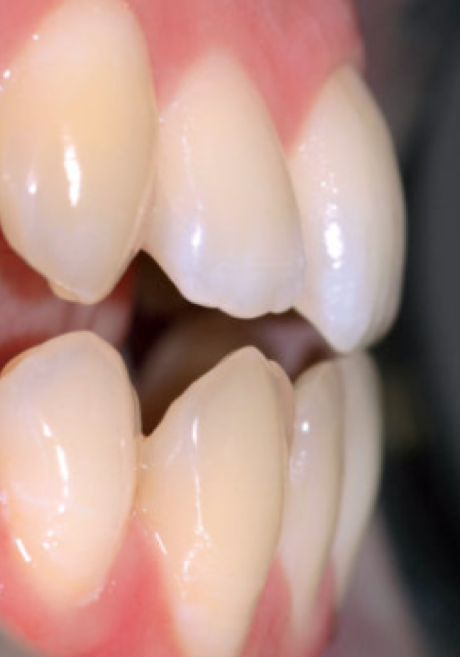
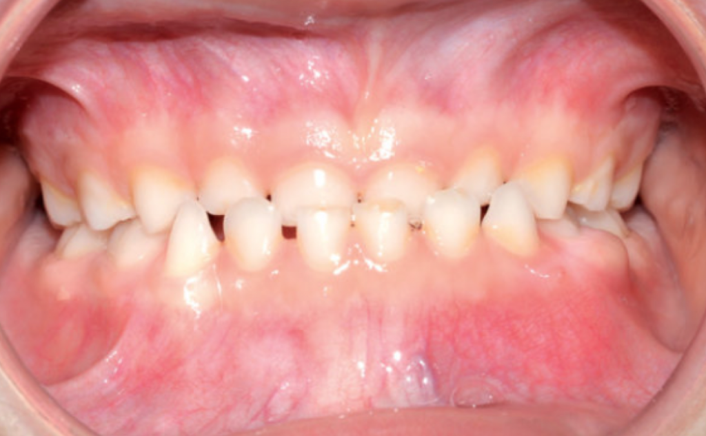
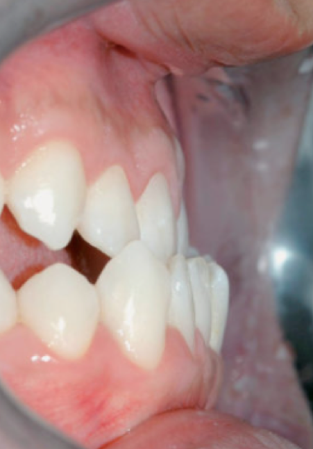
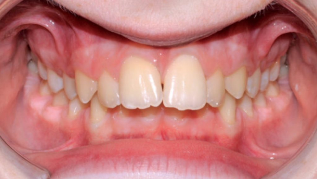
A crossbite represents a discrepancy in the vestibulo-lingual relationship of the upper and lower teeth. It can affect entire segments of the dental arch or individual teeth, or be mono- or bilateral. An anterior crossbite is present when the lower incisors occlude further forward than the upper incisors, resulting in a tooth relationship of complete inversion and decreased (or negative) overjet.

In dental anterior crossbite, one or more teeth are involved, a class I molar-canine relationship can be appreciated, and it may be due to abnormal axial tooth inclination. Functional anterior crossbite (or pseudo Class III) can be caused by the mandible tending to shift forward relative to the upper jaw as it closes. Skeletal anterior crossbite is characterized by a Class III molar and canine relationship in both centric occlusion and centric relationship. 

Posterior crossbite occurs when the upper posterior teeth occlude within the lower posterior teeth. It commonly occurs in a unilateral form with a functional displacement of the mandible toward the crossbite side. The etiology of posterior crossbite can include any combination of dental, skeletal, and neuromuscular functional components, but the most common cause is reduction in width of the upper arch. This reduction may be induced by vitiated habits such as thumb sucking, atypical swallowing, or upper airway obstruction caused by adenoid tissue or nasal allergies.

Bibliografia

1.      Sollenius O, Petrén S, Bondemark L. An RCT on clinical effectiveness and cost analysis of correction of unilateral posterior crossbite with functional shift in specialist and general dentistry. Eur J Orthod. 2020 Jan 27;42(1):44-51.

2.      Asiry MA, AlShahrani I. Prevalence of malocclusion among school children of Southern Saudi Arabia. J Orthod Sci. 2019;8:2.

3.      Yu X, Zhang H, Sun L, Pan J, Liu Y, Chen L. Prevalence of malocclusion and occlusal traits in the early mixed dentition in Shanghai, China. PeerJ. 2019;7:e6630

4.      Kutin, G. and R. R. Hawes. Posterior cross-bites in the deciduous and mixed dentitions. Am J Orthod 1969. 56:491–504.

5.      Melsen, B., K. Stensgaard, and J. Pedersen. Sucking habits and their influence on swallowing pattern and prevalence of malocclusion. Europ J Orthod 1979. 1:271–280.

6.      Linder-Aronson, S. Adenoids. Their effect on mode of breathing and nasal airflow and their relationship to characteristics of the facial skeleton and the dentition. Acta Otolaryngol 1970. 265:1–132.


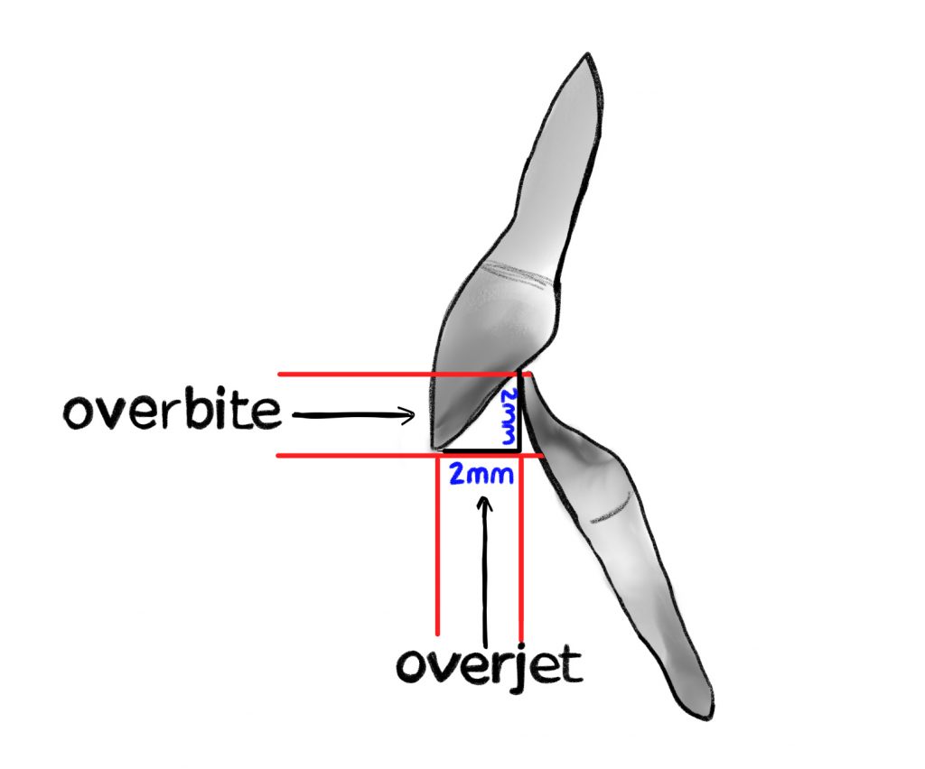
MALOCCLUSIONS ON THE VERTICAL PLANE

Vertical abnormalities can determine both alterations in facial aesthetics and functional problems and require early intervention in order to restore physiological environmental and functional conditions and promote physiological growth of the jaws.

An increase of the vertical dimension (open bite) can also determine alterations of the masticatory function, while an evident decrease (deep bite-deep bite) can support periodontal problems, in particular of the upper and lower incisors.
The open bite can be defined as a vertical incompetence of the arches determined by a dental and/or skeletal malposition that does not allow the occlusion of some dental elements with their respective antagonists. In particular, the anterior open bite represents the condition in which the crowns of the upper incisors are not able to overlap the crowns of the lower incisors, with decreased (or negative) overbite. 

The malpositioning can be a consequence of both an anomaly of the dento-alveolar district and an alteration of the development of the bones of the facial massive.
Etiologically, an anterior open bite can be related to skeletal, dental, and functional causes, generally in combination. Factors leading to the open bite condition include:

1. Spoiled habits (sucking on thumb or other fingers, atypical swallowing);

2.Low posture of the tongue at rest;

3. Respiratory problems related to allergies, adenoids and tonsils (due to the presence of excessive lymphatic tissue) and oral breathing. These patients, in order to breathe better, adopt an oral respiration that requires a low and anterior posture of the tongue to increase the airway volume;

4. Morphological-structural factors (macroglossia, tone and orientation of masticatory muscles);

5. Skeletal-structural factors (excess vertical anterior facial growth versus posterior vertical growth).
Deep bite is defined as increased vertical overlap between the upper and lower incisors and increased overbite. The reported prevalence of deep bite in orthodontic patients ranges from 11.8% to 36.7%. If left untreated, deep bite can lead to:

– Soft tissue trauma (palatal mucosa or vestibular gingiva of lower incisors) to the point of even causing gingival recessions;

– Temporo-mandibular disorders due to distalization of the mandible;

– Excessive abrasion of the anteriors (especially lower incisors);

– Impairment of the growth of the mandible
Genetic and acquired factors play a role in the development of a deep bite. The former include:

1. A horizontal skeletal growth pattern;

2. An over-eruption of the incisors;

3. An infra-occlusion of the molars.

Acquired factors include instead:

1. Lateral pushing of the tongue during swallowing;

2. Premature loss of deciduous teeth;

3. Abrasion of occlusal surfaces.

It is possible to distinguish between a skeletal and a dental deep bite. Very often the two conditions coexist together.

A skeletal deep bite is caused by an anti-clockwise rotation of the mandible, or a clockwise rotation of the maxilla, or a combination of both.

This type of patient typically has reduced anterior facial height and a horizontal growth pattern.

Dental deep bite is caused by over-eruption of the incisors or infra-occlusion of the molars, without the skeletal features seen for deep bite of skeletal origin.

The first condition is typical of skeletal class II with strong overjet and in these cases the lower incisors continue to erupt until they occlude with the palatal mucosa. Infra-occlusion of the molars is almost always caused by lateral lingual thrust or low, lateral tongue posture. Rarely, it may be caused by early loss of the posterior teeth.

Bibliografia

1.      Subtelny JD, Sakuda M. Open-bite: diagnosis and treatment. Am J Orthod 1964;50:337-358.

2.      Mucedero M, De Toffol L, Ballanti F, Donatelli M, Cozza P. Classificazione ezio-fisio-patologica del morso aperto. Mondo Ortod 2003;1:5-15.

3.      Maciel CT, Leite IC. Etiological aspects of anterior open bite and its implications to the oral functions. Pro Fono 2005;17(3):293-302.

4.      Cozza P, Mucedero M, Baccetti T, Franchi L. Early orthodontic treatment of skeletal open-bite malocclusion: a systematic review. Angle Orthod 2005 Sep;75(5):707-13.

5.      Jonsson T, Arnlaugsson S, Karlsson KO, Ragnarsson B, Arnarson EO, Magnusson TE. Orthodontic treatment experience and prevalence of malocclusion traits in an Icelandic adult population. Am J Orthod Dentofacial Orthop. 2007;131:8.e11–8.

6.      Borzabadi-Farahani A, Eslamipour F. Malocclusion and occlusal traits in an urban Iranian population. An epidemiological study of 11- to 14-year-old children. Eur J Orthod. 2009;31:477–84.

7.      Jackson S, Sandeler PJ. Fixed biteplanes for treatment of deep bite. J Clin Orthod. 1996; 30: 283-287.

8.      Philippe J. Treatment of deep bite with bonded bite planes. J Clin Orthod. 1996; 30: 396-400.